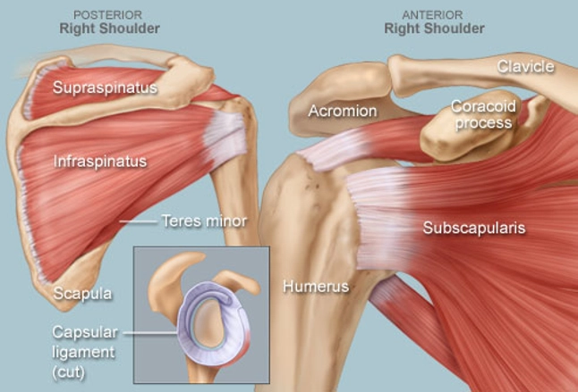
Shoulder Anatomy Diagram / Rotator cuff anatomy, anterior. | Download Scientific Diagram : Instead of your doctor simply saying that “the patient knee hurts”, he or she can say that “the patient’s knee hurts anterolaterally”.
Shoulder Anatomy Diagram / Rotator cuff anatomy, anterior. | Download Scientific Diagram : Instead of your doctor simply saying that "the patient knee hurts", he or she can say that "the patient's knee hurts anterolaterally".. These muscles form the outer shape of the shoulder and underarm. The joint capsule is lax, permitting greater mobility (particularly abduction). Numerous muscles help stabilize the three joints of. Starting with what is deepest, it goes: See full list on teachmeanatomy.info
Human anatomy diagrams show internal organs, cells, systems, conditions, symptoms and sickness information and/or tips for healthy living. These muscles form the outer shape of the shoulder and underarm. See full list on teachmeanatomy.info This is called the glenoid. Other important bones in the shoulder include:
Bones & Joints of the Shoulder | Orthopaedic - Hywel Williams from hywel-williams.co.uk Ebraheim's educational animated video describes muscle anatomy of the shoulder girdle and anatomy of the shoulder joint.anatomy of the shoulder muscles a. Numerous muscles help stabilize the three joints of. What are the names of the muscles in the shoulder? The muscles in the shoulder aid in a wide. What muscles attach to the shoulder? Anatomy termsallow us to describe the body and body motions more precisely. Anterior — the front of the shoulder 2. Inflammation of the bursa) can be a cause of shoulder pain.
To keep things simple, we can divide the shoulder into layers.
The glenoid is covered with smooth cartilage. It holds the tendon of the long head of the biceps in the intertubercular groove.] 4. Human anatomy diagrams show internal organs, cells, systems, conditions, symptoms and sickness information and/or tips for healthy living. In the shoulder joint, the ligaments play a key role in stabilising the bony structures. Anatomy termsallow us to describe the body and body motions more precisely. Instead of your doctor simply saying that "the patient knee hurts", he or she can say that "the patient's knee hurts anterolaterally". Starting with what is deepest, it goes: Nerves are like electrical wires that carry signals from the brain to the muscles to allow for movement of the shoulder. Where the rounded top of the arm bone (humerus) contacts the shoulder blade is called the glenohumeral joint. A bursa is a synovial fluid filled sac, which acts as a cushion between tendons and other joint structures. Mar 23, 2015 · the shoulder is a complex combination of bones and joints where many muscles act to provide the widest range of motion of any part of the body. The synovial membranelines the inner surface of the joint capsule, and produces synovial fluid to reduce friction between the articular surfaces. Below are some anatomic terms doctors use to describe location (as applied to the shoulder):
The shoulder joint is formed by the articulation of the head of the humerus with theglenoid cavity(or fossa) of the scapula. Thejoint capsuleis a fibrous sheath which encloses the structures of the joint. What are the different shoulder muscles? Starting with what is deepest, it goes: These two joints work together to allow the arm both to circumduct in a large circle and to rotate around its axis at the shoulder.
Shoulder Human Anatomy: Image, Function, Parts, and More from img.webmd.com Other important bones in the shoulder include: See full list on teachmeanatomy.info See full list on healthpages.org It supports the superior part of the joint capsule. See full list on healthpages.org The shoulder joint is formed by the articulation of the head of the humerus with theglenoid cavity(or fossa) of the scapula. The glenoid is covered with smooth cartilage. See full list on teachmeanatomy.info
Below are some anatomic terms doctors use to describe location (as applied to the shoulder):
What are the different shoulder muscles? The joint capsule is lax, permitting greater mobility (particularly abduction). They act to stabilise the anterior aspect of the joint. See full list on teachmeanatomy.info The synovial membranelines the inner surface of the joint capsule, and produces synovial fluid to reduce friction between the articular surfaces. Instead of your doctor simply saying that "the patient knee hurts", he or she can say that "the patient's knee hurts anterolaterally". Anterior — the front of the shoulder 2. What muscles attach to the shoulder? There are many nerves and blood vessels that supply the muscles and bones of the shoulder. The muscles in the shoulder aid in a wide. It extends from the anatomical neckof the humerus to the border or 'rim' of the glenoid fossa. The subacromial bursa reduces friction beneath the deltoid, promoting free motion of the rotator cuff tendons. Starting with what is deepest, it goes:
The shoulder joint is formed by the articulation of the head of the humerus with theglenoid cavity(or fossa) of the scapula. Proximal — located nearest to the point of attachment or reference, or center of the body 5.1. Numerous muscles help stabilize the three joints of. Ebraheim's educational animated video describes muscle anatomy of the shoulder girdle and anatomy of the shoulder joint.anatomy of the shoulder muscles a. See full list on teachmeanatomy.info
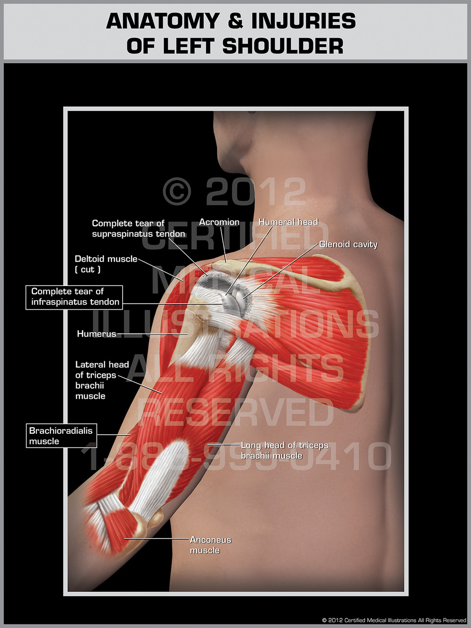
Anatomy & Injuries of Left Shoulder from cdn10.bigcommerce.com The shoulder joint is formed where the humerus (upper arm bone) fits into the scapula (shoulder blade), like a ball and socket. Thejoint capsuleis a fibrous sheath which encloses the structures of the joint. To reduce friction in the shoulder joint, several synovial bursaeare present. See full list on teachmeanatomy.info They also carry signals from the shoulder back to the brain about pain, pressure and temperature. The shoulder joint is formed by the articulation of the head of the humerus with theglenoid cavity(or fossa) of the scapula. This diagram depicts shoulder anatomy muscles diagram. Proximal — located nearest to the point of attachment or reference, or center of the body 5.1.
This is useful information, as the specific location of pain around body structures helps doctors and other health care providers to figure out what the cause of the patient's pain is.
Inflammation of the bursa) can be a cause of shoulder pain. See full list on teachmeanatomy.info What muscles attach to the shoulder? Anatomy termsallow us to describe the body and body motions more precisely. The bursae that are important clinically are: Jan 23, 2018 · the shoulder has about eight muscles that attach to the scapula, humerus, and clavicle. Where the rounded top of the arm bone (humerus) contacts the shoulder blade is called the glenohumeral joint. These two joints work together to allow the arm both to circumduct in a large circle and to rotate around its axis at the shoulder. It holds the tendon of the long head of the biceps in the intertubercular groove.] 4. Mar 23, 2015 · the shoulder is a complex combination of bones and joints where many muscles act to provide the widest range of motion of any part of the body. The muscles in the shoulder aid in a wide. This is useful information, as the specific location of pain around body structures helps doctors and other health care providers to figure out what the cause of the patient's pain is. Lateral — the side of the shoulder farthest from mid body 5.


0 Komentar